After a car accident, diagnosing injuries accurately is crucial for effective treatment and recovery. However, with various imaging options available, choosing the right diagnostic tool can be confusing. Two of the most commonly used imaging techniques are magnetic resonance imaging (MRI) and X-rays, each serving distinct purposes in the diagnostic process. Understanding the differences between these methods and when each is most beneficial can help you make informed decisions about your care.
In this blog, we will explore the key differences between MRI and X-ray imaging, highlighting their unique advantages and limitations. We’ll discuss which types of injuries are best suited for each imaging technique and offer guidance on how to determine the appropriate diagnostic tool for your specific needs. By the end, you’ll have a clearer understanding of how these technologies can aid in your recovery from a car accident.

Key Differences Between MRI and X-ray Imaging
Both MRI and X-ray imaging play crucial roles in diagnosing injuries after a car accident, but they offer distinct benefits and limitations based on their differing technologies and applications. Understanding these differences can help patients and healthcare providers choose the most suitable imaging technique for each unique situation.
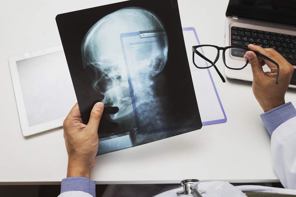
X-ray imaging is widely known for its ability to capture clear images of bones and detect structural abnormalities, such as fractures and dislocations. X-rays work by passing electromagnetic radiation through the body, with dense materials like bones absorbing more of the rays and appearing white on the resulting image. This method is fast, efficient, and relatively inexpensive, making it ideal for quickly assessing bone injuries. However, X-rays have limitations when it comes to soft tissues, as these appear as shadows or are not visible at all on the images. Therefore, while X-rays are excellent for diagnosing bone-related injuries, they are not suitable for evaluating soft tissue damage or internal organ injuries.
Magnetic resonance imaging (MRI), on the other hand, uses magnetic fields and radio waves to produce detailed images of the body’s internal structures. Unlike X-rays, MRIs excel at visualizing soft tissues, such as muscles, ligaments, and organs. This makes them invaluable for diagnosing injuries that involve soft tissue damage, such as torn ligaments, herniated discs, or internal bleeding. MRI scans provide highly detailed images, allowing healthcare providers to see fine details that might be missed with other imaging techniques. However, MRIs are more time-consuming and expensive compared to X-rays. Additionally, the powerful magnetic field generated during an MRI can be problematic for individuals with certain implants or medical devices, such as pacemakers, which can limit the use of this technology for some patients.
Both imaging techniques have their own set of advantages and limitations, and the choice between them depends on the specific injuries being evaluated. X-rays are quick and effective for bone-related injuries but fall short when it comes to soft tissue visualization. MRIs, while more comprehensive in their imaging capabilities, require more time, resources, and special considerations. Understanding these key differences is crucial for selecting the appropriate imaging method based on the nature of the injuries and the information needed for diagnosis and treatment.
Determining the Best Imaging Technique for Specific Injuries
Choosing the right imaging technique after a car accident depends largely on the nature of the injuries sustained. By understanding which types of injuries are best evaluated by MRI and which by X-ray, healthcare providers can ensure that patients receive the most effective and efficient care.
Injuries Best Suited for X-ray Imaging:
X-ray imaging is particularly effective for diagnosing bone-related injuries. This includes:
- Fractures: X-rays are the first line of imaging for suspected bone fractures. They can quickly reveal the presence, location, and pattern of fractures, helping to determine the appropriate treatment, whether it involves casting, splinting, or possibly surgery.
- Dislocations: X-rays can effectively show if and how bones have been displaced from their normal alignment within a joint, which is crucial for planning repositioning and stabilization treatments.
- Degenerative Bone Conditions: In cases where a patient has a pre-existing condition like osteoarthritis, X-rays can help assess the extent of joint or bone deterioration post-accident.
Injuries Best Suited for MRI Imaging:
MRI is invaluable for diagnosing conditions that involve soft tissues and non-bony structures. This includes:
- Soft Tissue Injuries: MRIs excel in visualizing soft tissue injuries, such as ligament tears, muscle strains, and tendon ruptures. These types of injuries are common in car accidents due to the force exerted on the body.
- Spinal Cord Injuries: MRI is critical for assessing any damage to the spinal cord or associated nerves, which might not be visible on an X-ray. It can detect issues like herniated discs, spinal stenosis, or nerve root compression.
- Brain Injuries: While CT scans are typically used for initial assessments of brain injuries, MRIs provide higher resolution images that are better at detecting more subtle brain injuries like concussions or diffuse axonal injuries.
Combining Both Modalities:
In some cases, both X-rays and MRIs are necessary to obtain a comprehensive understanding of the injury. For example, a patient might undergo X-ray imaging to assess a suspected fracture and then receive an MRI to evaluate additional soft tissue damage in the surrounding area.
The decision to use MRI or X-ray often depends on the initial assessment of the injury. X-rays may be used first for their speed and effectiveness in identifying obvious bone injuries, followed by MRI if more detailed imaging of soft tissues is required. Understanding the strengths and limitations of each imaging type ensures that patients are given the most appropriate and targeted diagnostic tests, leading to better treatment outcomes.
Determining the Appropriate Diagnostic Tool for Your Needs
Choosing the right imaging tool after a car accident is vital for accurate diagnosis and effective treatment. The decision should be based on several factors, including the nature of the injuries, symptoms, and the specific information needed for treatment planning. Here’s how to approach this decision-making process:
Consult with a Healthcare Provider: The first step in determining the appropriate diagnostic tool is consulting with a healthcare provider. Medical professionals are well-versed in the benefits and limitations of different imaging techniques and can recommend the best option based on your symptoms and medical history. A thorough evaluation by your doctor will guide the decision on whether an X-ray, MRI, or another form of imaging is necessary.
Consider the Type of Injury: The type of injury is a critical factor in choosing the right imaging tool. For example, if you suspect a bone-related injury such as a fracture or dislocation, an X-ray is often sufficient. However, if you’re experiencing symptoms that suggest soft tissue injuries, such as muscle tears or ligament damage, an MRI is likely more appropriate, as it provides detailed images of soft tissues.
Evaluate the Severity of Symptoms: The severity of your symptoms can also guide the choice of imaging. Severe or worsening pain, restricted movement, or neurological symptoms such as numbness or tingling may indicate more complex injuries that require detailed imaging. In such cases, an MRI might be warranted to provide a comprehensive view of the affected areas.
Factor in Time and Cost Considerations: Time and cost are practical considerations when choosing an imaging tool. X-rays are generally faster and more affordable than MRIs, making them a preferred choice for quick assessments or initial evaluations. If an X-ray reveals the need for further investigation, an MRI can then be considered. On the other hand, if time and budget are less of a concern, an MRI might be chosen upfront to gain a detailed understanding of the injuries.
Account for Personal Health Factors: Personal health factors, such as the presence of medical devices or claustrophobia, can affect the choice of imaging tool. MRIs, for instance, are not suitable for individuals with certain implants or medical devices, and they require patients to remain in a confined space for a prolonged period, which can be uncomfortable for those with claustrophobia. X-rays do not have these limitations, making them a more accessible option for some patients.
By considering these factors and working closely with healthcare providers, you can select the imaging tool that best fits your needs and supports your recovery.
Making the Right Choice for Your Car Accident Injury
Choosing the appropriate imaging tool, whether it’s an MRI or an X-ray, is crucial for accurately diagnosing injuries sustained in a car accident and ensuring effective treatment. Each imaging technique has its unique strengths and limitations, and the right choice depends on factors such as the type of injury, symptom severity, and individual health considerations.

At Doctor Wagner, we understand the importance of personalized care and use advanced imaging technologies to diagnose and treat car accident injuries effectively. Our team is dedicated to helping you navigate the recovery process, starting with accurate diagnostics and continuing with comprehensive treatment plans tailored to your needs.
If you’ve recently been in a car accident and are unsure about which imaging option is best for you, contact us today for expert advice and support. By making informed decisions about your diagnostic tools, you can set the foundation for a successful recovery and return to your normal activities with confidence.
